NOVEL TARGETED THERAPIES
Cancer doctors now know much more about how cancer cells function. New cancer therapies use this information to target cancer cell functions and stop them. Called targeted therapies, they can be more specific in stopping cancer cells from growing and may make other treatments work better. For example, some medicines work to prevent cancers from growing by preventing the growth of new blood vessels that would nourish the cancer. Other targeted therapies work more directly on cancer cells by blocking the action of molecules on the surface of cancer cells called growth factors.
RADIOSENSITIZERS
Any drug that can make tumor cells more sensitive to radiation is called a radiosensitizer. Combining radiation with radiosensitizers may allow doctors to kill more tumor cells. Some types of chemotherapy and some novel targeted therapies can act as radiosensitizers.
STEREOTACTIC RADIOTHERAPY
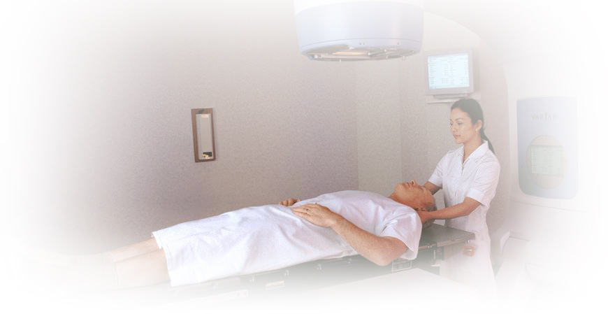
Stereotactic radiotherapy is a technique that allows your radiation oncologist to precisely focus beams of radiation to destroy certain types of tumors. Since the beam is so precise, your radiation oncologist may be able to spare more healthy tissue. This additional precision is achieved by using a very secure immobilization, such as a head frame used in the treatment of brain tumors. Stereotactic radiotherapy is frequently given in a single dose (sometimes called radiosurgery) although certain situations may require more than one dose. In addition to treating some cancers, radiosurgery can also be used to treat malformations in the brain’s blood vessels and certain noncancerous (benign) neurologic conditions. Sometimes a high dose of stereotactic radiotherapy can be focused upon a tumor outside the brain and given in a few treatments (typically three to eight). This form of treatment is called stereotactic body radiation therapy. IMAGE-GUIDED RADIATION THERAPY (IGRT) Radiation oncologists use image-guided radiation therapy, or IGRT, to help better deliver the radiation to the cancer since tumors can move between treatments due to diff erences in organ fi lling or movements while breathing. IGRT involves conformal radiation treatment guided by imaging, such as CT, ultrasound or X-rays, taken in the treatment room just before the patient is given the radiation treatment. Facts to Help Patients Make
an Informed Decision All patients first undergo a CT scan as part of the planning process. The imaging information from the CT scan is then transmitted to a computer in the treatment room to allow doctors to compare the earlier image with the images taken just before treatment. During IGRT, doctors compare these images to see if the treatment needs to be adjusted. This allows doctors to better target the cancer while avoiding nearby healthy tissue. In some cases, doctors will implant a tiny marker in or near the tumor to pinpoint it for IGRT.
THREE DIMENSIONAL CONFORMAL RADIATION THERAPY (3D-CRT)
Tumors are not regular — they come in diff erent shapes and sizes. Three-dimensional conformal radiation therapy, or 3D-CRT, uses computers and special imaging techniques to show the size, shape and location of the tumor. Computer assisted tomography (CT or CAT scans) or magnetic resonance imaging (MR or MRI scans) are used to create detailed, three-dimensional representations of the tumor and surrounding organs. Your radiation oncologist can then precisely tailor the radiation beams to the size and shape of your tumor with multileaf collimators (see picture, right) or custom fabricated field shaping blocks. Because the radiation beams are very precisely directed, nearby normal tissue receives less radiation and is able to heal quickly. |